Top this week

most clinically relevant
Interventional (IR)Musculoskeletal (MSK)ResearchTrainee

Osteoporosis medication after vertebral augmentation linked to 6-fold lower refracture risk

Only one-third of patients received osteoporosis medication after vertebral augmentation. The incidence of new vertebral fractures fell from 0.519 to 0.076 per person-year after starting medical management (incidence rate ratio 6.79, 95% CI 3.37-15.51), a 6-fold decreased risk.

Journal of vascular and interventional radiology : JVIR
Chest / ThoracicAI / InformaticsResearch

Integrated CT Biomarker Model Boosts COPD Detection in Lung Cancer Screening

Integrated model combining emphysema, Pi10, airway branch count, and clinical data detected undiagnosed COPD with AUC 0.83 (95% CI 0.80-0.86) in 5,014 lung screening participants, up from 0.69 for emphysema alone, reducing confirmatory spirometry referrals from 41% to 34%.

Chest

Latest

3181 stories
Musculoskeletal (MSK)EducationTrainee

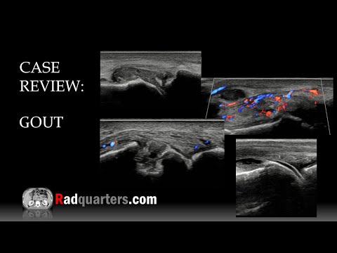
Sonographic Recognition of Gout: Core Imaging Pearls

This lecture reviews the ultrasound features of gout, including the double contour sign and tophi, in a condition that typically presents as acute monoarthritis of the first MTP joint in men >40 with hyperuricemia.

Radquarters (YouTube)
Neuro / Head & NeckEducationTrainee

Stroke complications and mimics: A video guide for radiologists

A new educational video reviews stroke complications like hemorrhagic conversion—masslike hemorrhage >1 cm with mass effect—and CT perfusion mimics, noting that patients with petechial hemorrhage may safely continue anticoagulation.

LearnNeuroradiology (YouTube)
Neuro / Head & NeckEmergencyEducationTrainee

Stroke Imaging Case: Distal Basilar Occlusion with Vertebral Irregularity on CTA and DSA

A 50-year-old with altered mental status had a non-contrast head CT reported as normal. CT angiogram revealed a focal occlusion of the distal basilar artery extending into the right P1 segment, with non-opacification of the proximal left PCA and left vertebral artery. DSA confir…

LearnNeuroradiology (YouTube)
Neuro / Head & NeckPediatricEducationTrainee

Medullary Cap Dysplasia with Spinal Dysraphism: A Rare Association

Eurorad teaching case presents medullary cap dysplasia, a rare congenital spinal cord anomaly, and its association with spinal dysraphism. Recognizing this entity on MRI can prevent misdiagnosis.

Eurorad (ESR teaching cases)
GeneralEducationTrainee

Quick Video Explains Hounsfield Unit Scale in CT Imaging

A short educational video from Navigating Radiology provides a simple explanation of Hounsfield units (HU), the standardized scale used in CT to quantify tissue density. Aimed at radiology residents and medical students, it covers how water is 0 HU and why the scale matters for…

Navigating Radiology (YouTube)
Neuro / Head & NeckEducationTrainee

Stroke Imaging Case 2: Left MCA Occlusion with Core Infarct and Penumbra

Educational stroke imaging case: 72yo with right weakness, CT shows left insular hypodensity, CTP small core with large penumbra, DSA confirms distal M1/M2 occlusion. Successful thrombectomy. Part of a 4-case intro series.

LearnNeuroradiology (YouTube)
Neuro / Head & NeckEducationTrainee

Short Video Review of Brain Arterial Territories

Educational short reviews the arterial territories of the brain: ACA (anterior frontal), MCA (lateral frontal, parietal, temporal), PCA (medial parietal, occipital, temporal), and SCA (superior, medial cerebellum). A quick reference for recognizing vascular patterns on imaging.

LearnNeuroradiology (YouTube)
BreastResearchTrainee

Higher-Than-Expected PPV of BI-RADS 4A Lesions in Indian Tertiary Centre

In a retrospective study of 230 women with BI-RADS 4 lesions, positive predictive value (PPV) was 26.9%. Subcategory PPVs: 4A 18.7% (exceeding typical 2-10%), 4B 27.6%, 4C 50%. History of breast cancer increased malignancy risk (34.9% vs 21.5%, p=0.001).

Reporting systems & Fleischner (PubMed)
GeneralEducationTrainee

CT Grayscale Explained: What Those Shades of Gray Actually Mean

New educational short video from Navigating Radiology breaks down how different shades of gray on CT correspond to tissue densities (e.g., bone, fluid, fat, air), helping learners quickly interpret scans.

Navigating Radiology (YouTube)
Neuro / Head & NeckEmergencyEducationTrainee

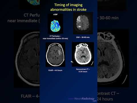
Timing of imaging findings after a stroke: from CT perfusion to postcontrast enhancement

Key stroke imaging timelines: CT perfusion detects abnormalities near-immediately (<30 min). DWI turns positive at 30-60 min. FLAIR changes appear at 4-6 h. Noncontrast CT becomes abnormal at 6-24 h. Postcontrast enhancement is seen at 2-3 days. A quick reference for image inter…

LearnNeuroradiology (YouTube)
GeneralEducationTrainee

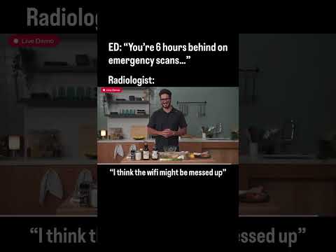
Using Humor to Teach the Realities of Radiology Workflow Frustrations

A short, humorous educational vignette highlights common, non-clinical pitfalls in radiology workflow—like network and PACS failures—that disrupt reading efficiency. It serves as lighthearted, relatable training for residents and trainees navigating the daily technical frustrati…

Navigating Radiology (YouTube)
Neuro / Head & NeckEducationTrainee

MRI clues for perineural and leptomeningeal tumor spread

Contrast-enhanced MRI is highly sensitive for detecting perineural and leptomeningeal tumor spread. This video with Dr. Katie Bailey shows key imaging features and differences, using case examples.

LearnNeuroradiology (YouTube)
Neuro / Head & NeckPediatricEducationTrainee

MRI Teaching Case: Rings and Blooming in Coats Plus Syndrome

This Eurorad teaching case highlights the MRI features of Coats plus syndrome, emphasizing 'rings and blooming' as diagnostic clues. It serves as a visual walk-through for radiologists encountering this rare neurocutaneous disorder.

Eurorad (ESR teaching cases)
Musculoskeletal (MSK)EducationTrainee

Knee MRI Anatomy & Approach: A Comprehensive Video Guide

Free comprehensive video tutorial on knee MRI from Navigating Radiology, covering sequences, labeled anatomy, and a systematic approach to each structure. Includes case-based modules with AI hints and feedback to learn common pathologies.

Navigating Radiology (YouTube)
Body / AbdominalEmergencyGeneralEducationTrainee

Ultrasound essentials for detecting common bile duct stones

On ultrasound, common bile duct (CBD) stones may lack posterior shadowing; use twinkling artifact and a dilated CBD (>6 mm, +1 mm per decade >60 years) as key clues. Seen in up to 15% of patients with gallstones, complications include cholangitis and pancreatitis.

Radquarters (YouTube)
Body / AbdominalGeneralEducationTrainee

Small Bowel Diverticulitis: Radiologic Teaching Pearl

Brief educational video reviewing the typical imaging features of small bowel diverticulitis on CT and MRI, with emphasis on differentiating it from other acute abdominal conditions.

Radquarters (YouTube)
Body / AbdominalEducationTrainee

Contrast-Enhanced US of Focal Nodular Hyperplasia: A Leave-Alone Lesion

Focal nodular hyperplasia (FNH) is a benign liver tumor with no malignant potential, requiring no surgery—a true 'leave alone' lesion. CEUS aids diagnosis. Most common in women aged 20-50, with an 8:1 female-to-male ratio, and no link to oral contraceptives.

Radquarters (YouTube)
Neuro / Head & NeckEducationTrainee

Short Video Series Distills Neuroradiology Concepts: Prolactinoma

A new 'Brain Bites' video educates on prolactinomas, a type of pituitary adenoma, the most common sellar mass. This installment, presented by a medical student, offers a quick, digestible guide to the imaging features of this common brain tumor.

LearnNeuroradiology (YouTube)
Neuro / Head & NeckEducationTrainee

Brain MRI: Blind Spots and Pearls for Radiologists

This Navigating Radiology course teaches radiologists to identify commonly missed brain MRI findings—blind spots and nuanced anatomy—to avoid missing important incidental findings in daily practice.

Navigating Radiology (YouTube)
Neuro / Head & NeckGeneralEducationTrainee

Video Guide: How to Read Routine Brain MRI Sequences

A new educational video from Navigating Radiology provides a foundational approach to reading brain MRIs, covering routine sequences, normal anatomy, and patterns of abnormal enhancement. The course aims to help learners 'look at studies ASAP' and is followed by a second video a…

Navigating Radiology (YouTube)
Musculoskeletal (MSK)EducationTrainee

Ultrasound Features of Intersection Syndrome at the Forearm

Intersection syndrome is a friction tenosynovitis where the first extensor compartment crosses the second, 4–8 cm proximal to Lister's tubercle. This video reviews its ultrasound appearance.

Radquarters (YouTube)
Neuro / Head & NeckEducationTrainee

Neuroacanthocytosis Syndrome: Eurorad Teaching Case

Eurorad teaching case presents neuroacanthocytosis syndrome (chorea-acanthocytosis), a rare neurodegenerative disorder characterized by chorea and acanthocytes on peripheral smear.

Eurorad (ESR teaching cases)
NewerPage 93 of 107Older