BreastEducationTrainee
Breast anatomy for imaging interpretation
Radiology Tutorials (YouTube)today
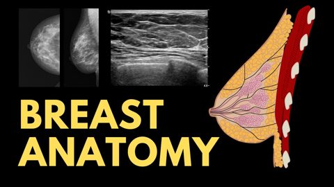
This lecture breaks down breast anatomy with a practical focus on imaging: lobes, lobules, ducts, skin/muscle relations, Cooper's ligaments, blood supply, and lymphatic drainage — all correlated with what we see on mammography and ultrasound.
- Covers lobar and ductal anatomy and how they appear on mammography and ultrasound.
- Details suspensory ligaments (Cooper's) and their role in imaging interpretation.
- Lymphatic drainage patterns are highlighted for accurate lesion localization.
Automated summary
RadPigeon summaries are original and for information only. They are not clinical advice.