Body / AbdominalEducationTrainee
Serous Cystadenoma on Axial CT: A Teaching Case
CTisus (YouTube)Jul 9
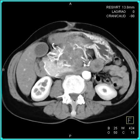
CTisus teaching case illustrates axial CT features of pancreatic serous cystadenoma: microcystic (honeycomb) appearance, central scar, and benign behavior. Key for distinguishing from mucinous lesions.
- Short educational video from CTisus (YouTube) demonstrating a classic case of serous cystadenoma.
- Highlights the microcystic architecture and central calcified scar on axial CT.
- Ideal for trainees learning pancreatic cystic lesion characterization.
Automated summary
RadPigeon summaries are original and for information only. They are not clinical advice.